

Acute severe mitral regurgitation (MR) and aortic regurgitation (AR) in the critical care setting offer unique challenges, presenting as decompensated heart failure or cardiogenic shock that responds poorly to standard medical therapies. Decompensated valvular heart disease accounts for approximately 8% of cardiac ICU admissions, with up to 40% in-hospital mortality when complicated by cardiogenic shock.1 Acute MR is most often due to chordae tendineae rupture, papillary muscle rupture, or infective endocarditis. Acute severe AR, caused primarily by infective endocarditis, aortic dissection, or trauma, carries similarly high mortality from pulmonary edema, arrhythmias, and circulatory collapse and nearly always requires emergent surgery.2 Severe aortic stenosis (AS) is one of the most common chronic decompensated valvular diseases in patients who are critically ill.
Valvular dysfunction presents a diagnostic challenge in patients who are critically ill, particularly those with cardiogenic shock. Physical examination may be limited, as murmurs may be relatively “silent” due to rapid/early equalization of pressures across the dysfunctional valve. Point-of-care echocardiography, transthoracic echocardiography (TTE), and transesophageal echocardiography (TEE) thus become valuable tools for bedside or intraoperative assessment of valvular function in patients who are rapidly decompensating. Echo allows assessment of valvular morphology; color flow and continuous wave (CW) Doppler offer assessment and visualization of the regurgitant jet, jet area, and vena contracta (VC). A denser CW Doppler signal indicates larger regurgitant flow. However, in acute severe AR, left ventricular (LV) diastolic pressure rises rapidly due to both atrial filling and regurgitant volume resulting in the regurgitant jet being shorter and with lower velocity.

Severe AR is marked by large central jets or variable eccentric jets, holodiastolic flow reversal, VC >6 mm, and regurgitant fraction ≥50%.3 Severe MR on TTE shows holosystolic CW jet, large central jet >50% of left atrium, VC ≥7 mm, and regurgitant fraction ≥50%. Severe AS on TEE demonstrates AS jet velocity >4 m/s, mean pressure gradient >40 mm Hg, and effective orifice area <1 cm.2 Concurrent assessment of ejection fraction is imperative to guide the management of valvular dysfunction.4 Additionally, detailed phenotyping of cardiogenic shock in patients with a pulmonary artery catheter can result in better outcomes.5
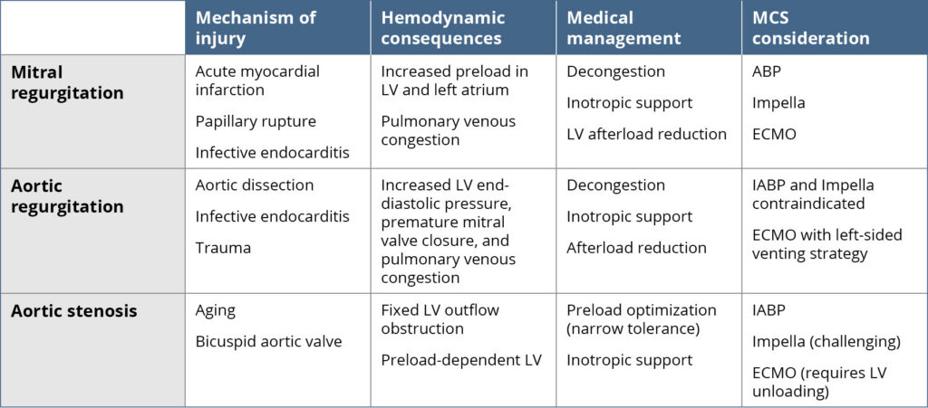
Medical therapy in acute severe MR and AR is primarily a temporizing bridge to definitive surgical or transcatheter intervention.1 For acute MR, afterload reduction with sodium nitroprusside or nicardipine is the first-line treatment in patients who are normotensive, reducing regurgitant volume by redirecting flow into the aorta.6 In acute AR, afterload reduction can allow temporary stabilization. In patients who are hypotensive, vasopressors including norepinephrine, epinephrine, or dopamine are reasonable choices, as they provide inotropy as well; purely vasoconstrictive agents like phenylephrine should be avoided, as they increase afterload and worsen MR.1 In acute AR, β-blockers should be avoided because blocking compensatory tachycardia prolongs diastole and worsens regurgitant volume.1,6 Diuretics address pulmonary congestion in both conditions. Positive pressure ventilation can facilitate gas exchange while reducing LV preload and afterload. Intubation in acute severe MR and AR carries high risk of cardiovascular collapse; etomidate or ketamine is the preferred induction agent to minimize hemodynamic compromise, with vasopressors at bedside prior to induction.1,7 Once intubated, fully supported ventilator modes with tidal volumes of 6 to 8 mL/kg ideal body weight and moderate positive end-expiratory pressure (PEEP) should be used, as PEEP reduces LV afterload and regurgitant volume in acute MR. Although, caution is warranted in concomitant right ventricular dysfunction, where lower PEEP may be necessary.1,8,9

Mechanical circulatory support (MCS) can be a useful bridge to definitive valve therapy when medical stabilization fails, but device choice must match valve physiology.1 In severe AS, intra-aortic balloon pump (IABP) offers only modest support, and Impella may be technically challenging or ineffective across a fixed stenosis; venoarterial extracorporeal membrane oxygenation (ECMO) can rescue perfusion but may require LV unloading. In acute severe AR, MCS is more constrained because IABP can worsen regurgitation and Impella requires crossing an incompetent aortic valve; if ECMO is used, it often needs an unloading strategy such as left atrial venoarterial ECMO or another left-sided venting approach.10 In severe MR—especially when shock persists despite diuresis, vasodilators, and inotropes—IABP, Impella, or ECMO can be considered as a short-term bridge to surgery or transcatheter intervention, ideally guided by invasive hemodynamics and echo reassessment.
Table 1
References
1. Miller PE, Senman BC, Gage A, et al. Acute decompensated valvular disease in the intensive care unit. JACC Adv. 2024;3(12):101402. doi:10.1016/j.jacadv.2024.101402
2. Roberts WC, Ko JM, Moore TR, Jones WH III. Causes of pure aortic regurgitation in patients having isolated aortic valve replacement at a single US tertiary hospital (1993 to 2005). Circulation. 2006;114(5):422-429. doi:10.1161/CIRCULATIONAHA.106.622761
3. Tsampasian V, Victor K, Bhattacharyya S, Oxborough D, Ring L. Echocardiographic assessment of aortic regurgitation: a narrative review. Echo Res Pract. 2024;11(1):1. doi:10.1186/s44156-023-00036-7
4. Vahanian A, Beyersdorf F, Praz F, et al. 2021 ESC/EACTS guidelines for the management of valvular heart disease. Eur Heart J. 2022;43(7):561-632. doi:10.1093/eurheartj/ehab395
5. Garan AR, Kanwar M, Thayer KL, et al. Complete hemodynamic profiling with pulmonary artery catheters in cardiogenic shock is associated with lower in-hospital mortality. JACC Heart Fail. 2020;8(11):903-913. doi:10.1016/j.jchf.2020.08.012
6. Otto CM, Nishimura RA, Bonow RO, et al. 2020 ACC/AHA guideline for the management of patients with valvular heart disease: a report of the American College of Cardiology/American Heart Association joint committee on clinical practice guidelines. Circulation. 2021;143(5):e72-e227. doi:10.1161/CIR.0000000000000923
7. Kotani Y, Russotto V. Induction agents for tracheal intubation in critically ill patients. Crit Care Med. 2025;53(1):e173-e181. doi:10.1097/CCM.0000000000006506
8. Fordyce CB, Katz JN, Alviar CL, et al. Prevention of complications in the cardiac intensive care unit: a scientific statement from the American Heart Association. Circulation. 2020;142(22):e379-e406. doi:10.1161/CIR.0000000000000909
9. Alviar CL, Miller PE, McAreavey D, et al. Positive pressure ventilation in the cardiac intensive care unit. J Am Coll Cardiol. 2018;72(13):1532-1553. doi:10.1016/j.jacc.2018.06.074
10. Santana JM, Dalia AA, Newton M, et al. Mechanical circulatory support options in patients with aortic valve pathology. J Cardiothorac Vasc Anesth. 2022;36(8 Pt B):3318-3326. doi:10.1053/j.jvca.2022.04.010
