

The latest American Society of Echocardiography (ASE) guidelines incorporate the updated World Symposium on Pulmonary Hypertension definition (mean pulmonary arterial pressure [mPAP] >20 mm Hg and pulmonary vascular resistance [PVR] >2 Wood units) and emphasize three pillars of right-heart assessment: structure, function, and hemodynamics.1,2
Structure
The guidelines recommend assessing the right ventricle (RV) for chronic pressure and volume loading, including RV diameter, right atrial (RA) size, RV wall thickness, and left ventricular (LV) eccentricity index (LVEI). Reports should include at least one quantitative RV size measurement, preferably RV basal diameter obtained via the apical four-chamber view. RA enlargement is a marker of chronic RV dysfunction, with each 1-cm2 RA area increase conferring a 6% morbidity risk increase.2

RV thickening reflects pressure overload but is nonspecific. LVEI is calculated as LV diameter parallel to the interventricular septum (IVS)/LV diameter perpendicular to the IVS. Progressive septal flattening and leftward apical displacement assessed by LVEI indicate advanced RV maladaptation and ventricular interdependence.
Numbers to know
- Normal RV basal diameter: <4.1 cm; severe dilatation: >4.9 cm
- Normal RA area: <19 cm2; severe enlargement: >24 cm2
- RA volume index: normal <30 mL/m2; severe dilation >41 mL/m2
- Normal RV wall thickness: <0.5 cm; severe hypertrophy: >0.9 cm
- LVEI >1 suggests volume overload
- An abnormal value in diastole alone suggests volume overload.
- Abnormalities in both systole and diastole suggest pressure with or without volume overload.

Function
Structural measures identify RV remodeling; functional measures assess afterload adaptation. The initial RV response to PH is compensatory hypertrophy with preserved function; progressive pressure overload subsequently leads to RV dilation, reduced contractility, adverse ventricular interdependence, and RV-PA uncoupling.
Assessment of RV function relies on four primary measures: Tricuspid annular plane systolic excursion (TAPSE), tissue Doppler S′ velocity, fractional area change (FAC), and strain (RV free-wall strain [RVFWS] and RV global longitudinal strain).
TAPSE can be misleading in severe tricuspid regurgitation (TR), altered RV geometry, pacing/catheter presence, postoperative states, or poor alignment. If TAPSE looks abnormal, corroborate it with tissue Doppler S′ velocity (movement of the basal RV free wall during systole), FAC of the RV, and RVFWS (useful for detecting subclinical disease).
Numbers to know
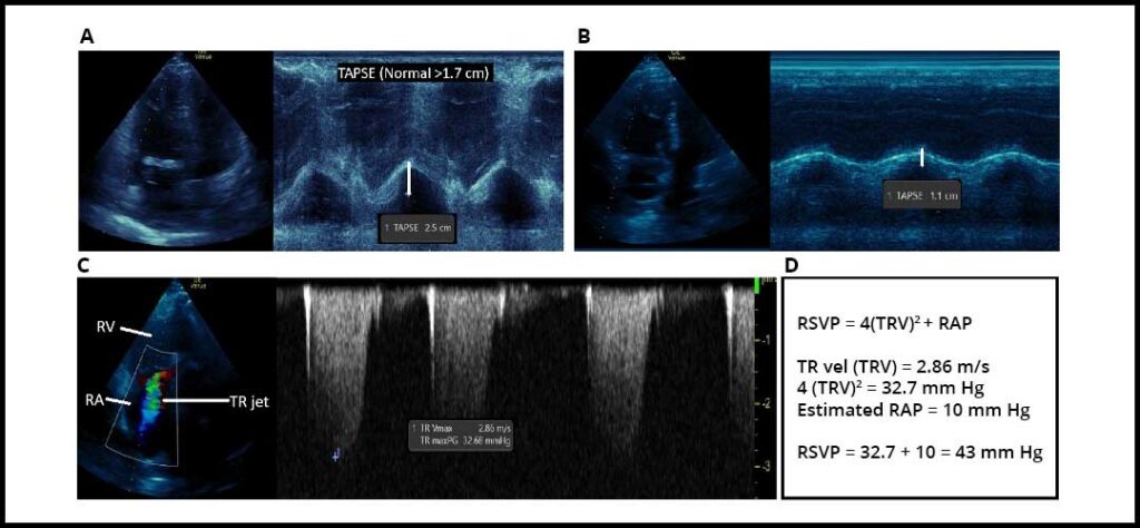
- Normal TAPSE: >1.7 cm; severely reduced: ≤1 cm (Figure 1); TAPSE should not be interpreted in isolation as evidence of RV dysfunction
- Normal tissue Doppler S′ velocity: >9.5 cm/s; severely reduced: ≤5.0 cm/s
- Normal fractional area change (FAC): >35%; severely reduced: ≤22%
- Normal RV free-wall strain (RVFWS): more negative than -20%; severe reduction: less negative than -11%
Hemodynamics
Structural and functional findings characterize the RV performance while Doppler-derived measures quantify hemodynamic load. TR velocity (TRV) and RV systolic pressure (RVSP) calculations help evaluate the probability of PH. Still, they are affected by RA pressure (RAP), cardiac output, pulmonary capillary wedge pressure (PCWP), and PVR. In the absence of a gradient across the pulmonic valve or RV outflow tract (RVOT), PA systolic pressure (PASP) is equal to RVSP. RVOT acceleration time shortens as pulmonary vascular impedance rises.
No single Doppler-derived pressure reliably reflects disease severity, and a high RVSP with a relatively preserved RV may pose less risk of decompensation than a modest RVSP with a severely failing RV. RV-PA coupling is assessed and expressed as the ratio of TAPSE/PASP. LV filling pressure is estimated using the mitral E/e′ obtained during diastolic assessment and can serve as a noninvasive PCWP correlate.
Numbers to know
- Normal RVSP: ≤34 mm Hg
- TRV below 2.8 m/s is reassuring; however, TRV ≥2.8 m/s, accompanied by at least two supportive echocardiographic signs, suggests PH
- RAP should be reported as a single number based on inferior vena cava (IVC) diameter and collapse
- 3 mm Hg when normal and collapsing
- 8 mm Hg if indeterminate
- 15 mm Hg when dilated and noncollapsing
- 20 mm Hg is reserved for marked dilation with evidence of stagnant flow
- RVOT AccT is normal above 105 ms
- TAPSE/PASP (coupling evaluation):
- Normal: 0.5-0.7
- A 0.3-0.4 or lower ratio is associated with increased mortality
- A rest-stress mitral E/e′ >12 ratio suggests a postcapillary component
Summary
A PH-focused echocardiogram should routinely report TRV/RVSP, RA size, RV size, septal configuration, and RV systolic function, while noting any pericardial effusion as a marker of advanced disease. The key clinical question is whether the RV is adapting to its afterload or becoming progressively dilated and uncoupled. Although right heart catheterization remains essential for definitive hemodynamic classification, the 2025 ASE guidelines provide a critical framework for standardized, longitudinal right-heart assessment.
References
1. Kovacs G, Bartolome S, Denton CP, et al. Definition, classification and diagnosis of pulmonary hypertension. Eur Respir J. 2024;64(4):2401324. doi:10.1183/13993003.01324-2024
2. Mukherjee M, Rudski LG, Addetia K, et al. Guidelines for the echocardiographic assessment of the right heart in adults and special considerations in pulmonary hypertension: recommendations from the American Society of Echocardiography. J Am Soc Echocardiogr. 2025;38(3):141-186. doi:10.1016/j.echo.2025.01.006
