

Your hospitalist colleague transfers a patient to the ICU because of worsening shortness of breath. The patient is a 60-year-old man who was admitted to the medical ward the previous day with pneumonia. On examination, he is in severe respiratory distress, and you determine that endotracheal intubation is necessary. A review of his home medications reveals that he is taking a glucagon-like peptide-1 (GLP-1) receptor agonist. Although the bedside nurse reports that his last meal was more than eight hours ago, you are now concerned about his risk of aspiration.
This scenario is becoming increasingly common in the ICU as the use of GLP-1 receptor agonists continues to rise.
Fasting duration alone is a poor predictor of gastric contents and aspiration risk, particularly in patients receiving GLP-1 receptor agonists or opioids, or those with gastroparesis or neuromuscular disorders.1 Furthermore, ICU clinicians frequently encounter situations requiring emergency intubation or circumstances in which fasting status cannot be reliably confirmed.

Gastric point-of-care ultrasound (POCUS) is a reliable bedside technique for assessing gastric contents and estimating aspiration risk.2,3 Already widely adopted in anesthesiology, gastric ultrasound is increasingly being incorporated into emergency medicine and critical care practice. Although most validation studies have been performed in elective surgical patients, emerging evidence suggests that gastric ultrasound is both feasible and clinically useful in patients who are critically ill.4 Beyond preintubation assessment, it also has important applications before extubation.
Whenever feasible, gastric ultrasound images should be obtained in both the supine and right lateral decubitus (RLD) positions.5,6 A practical approach is to place a curvilinear transducer in the sagittal orientation just inferior to the xiphoid process and gradually move caudally while identifying the rectus abdominis muscles, left lobe of the liver, and stomach, sweeping the transducer from left to right. The antrum can be identified by its typical location under the left lobe of the liver and its characteristic thick muscularis (Figure 1A).
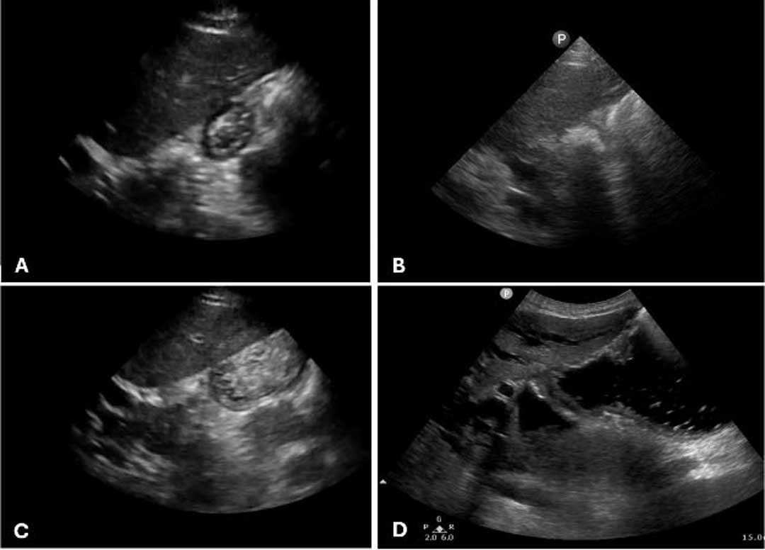
To prevent inadvertent imaging of the pylorus, the aorta should be visualized in the distance before reducing the depth to focus on the antrum. The antrum should be observed for at least 15 to 20 seconds, as gastric peristalsis can significantly alter its appearance.
Image interpretation is relatively straightforward when the stomach is either empty (Figure 1A) (low aspiration risk) or contains solid or thick particulate material (Figure 1B), which appears hyperechoic and indicates a high aspiration risk. When the antrum contains clear fluid, producing a hypoechoic or anechoic appearance, interpretation becomes more nuanced.
Figure 1C shows a small amount of fluid with air bubbles; Figure 1D shows a distended stomach with clear fluid. In these cases, gastric volume can be estimated by measuring the cross-sectional area of the gastric antrum and applying validated predictive equations.7 The estimated gastric volume can then be compared with proposed aspiration risk thresholds, generally ranging from 0.8 to 1.5 mL/kg.8,9
A qualitative grading system has also been described. Grade 0 indicates no fluid visible in either the supine or RLD position; Grade 1 indicates fluid visible only in the RLD position; and Grade 2 indicates fluid visible in both positions. Grades 0 and 1 are generally considered low risk for aspiration, whereas Grade 2 is associated with a higher aspiration risk.2
The findings from gastric ultrasound can directly influence airway management. Depending on the urgency of the clinical situation and the ultrasound findings, clinicians may elect to delay intubation when appropriate, decompress the stomach with a nasogastric tube before induction, or prepare for a high-risk intubation using strategies designed to minimize aspiration.
Beyond preintubation assessment, gastric ultrasound has several additional applications in the ICU, including confirmation of gastric tube placement, monitoring of gastric emptying and enteral feeding tolerance, and assessment of gastric volume before extubation.10,11 The examination is relatively simple, can be completed within a few minutes, and can be readily taught to physicians and appropriately trained nursing staff.
Despite its advantages, clinicians should recognize several limitations of gastric ultrasound. Examination in the supine position alone may fail to detect gastric contents that redistribute into the antrum only in the RLD position, potentially resulting in false reassurance. Mistaking adjacent vascular structures for the gastric antrum can be avoided by using color Doppler imaging. Excessive intragastric gas may obscure visualization of the antrum. In addition, previous gastric surgery, large hiatal hernias, obesity, or congenital gastric anomalies may limit image acquisition and interpretation.
As gastric ultrasound becomes more widely adopted in critical care, it has the potential to become an invaluable bedside tool for individualized aspiration risk assessment and procedural decision-making. In an era of increasing GLP-1 receptor agonist use and frequent emergency airway interventions, gastric POCUS provides clinically actionable information that fasting history alone cannot.
References
1. Nguyen M, Drihem A, Berthoud V, et al. Fasting does not guarantee empty stomach in the intensive care unit: a prospective ultrasonographic evaluation (The NUTRIGUS study). Anaesth Crit Care Pain Med. 2021;40(6):100975. doi:10.1016/j.accpm.2021.100975
2. Kruisselbrink R, Gharapetian A, Chaparro LE, et al. Diagnostic accuracy of point-of-care gastric ultrasound. Anesth Analg. 2019;128(1):89-95. doi:10.1213/ANE.0000000000003372
3. Ruiz Ávila HA, Espinosa Almanza CJ, Fuentes Barreiro CY. Inter-observer and intra-observer variability in ultrasound assessment of gastric content and volume in critically ill patients receiving enteral nutrition. Ultrasound J. 2023;15(1):14. doi:10.1186/s13089-023-00312-x
4. Hamada SR, Garcon P, Ronot M, Kerever S, Paugam-Burtz C, Mantz J. Ultrasound assessment of gastric volume in critically ill patients. Intensive Care Med. 2014;40(7):965-972. doi:10.1007/s00134-014-3320-x
5. Foster B, Chen J, Tsui BCH. A stepwise approach to locating the antrum during gastric ultrasound. J Cardiothorac Vasc Anesth. 2023;37(3):498-499. doi:10.1053/j.jvca.2022.11.030
6. Holtan-Hartwig I, Johnsen LR, Dahl V, Haidl F. Preoperative gastric ultrasound in surgical patients who undergo rapid sequence induction intubation. Trends Anaesth Crit Care. 2021;38:30-35. doi:10.1016/j.tacc.2021.04.005
7. Rocha CATD, Kamada LMK, Andrade Filho PH, Villaverde IA, Shiro JYB, Silva Junior JMD. Ultrasonographic evaluation of gastric content and volume: a systematic review. Rev Assoc Med Bras (1992). 2020;66(12):1725-1730. doi:10.1590/1806-9282.66.12.1725
8. Bouvet L, Mazoit JX, Chassard D, Allaouchiche B, Boselli E, Benhamou D. Clinical assessment of the ultrasonographic measurement of antral area for estimating preoperative gastric content and volume. Anesthesiology. 2011;114(5):1086-1092. doi:10.1097/ALN.0b013e31820dee48
9. Asokan R, Bhardwaj BB, Agrawal N, et al. Point-of-care gastric ultrasound to predict aspiration in patients undergoing urgent endotracheal intubation in the emergency department. BMC Emerg Med. 2023;23(1):111. doi:10.1186/s12873-023-00881-z
10. Brotfain E, Erblat A, Luft P, et al. Nurse-performed ultrasound assessment of gastric residual volume and enteral nasogastric tube placement in the general intensive care unit. Intensive Crit Care Nurs. 2022;69:103183. doi:10.1016/j.iccn.2021.103183
11. Nguyen M, Ouharani A, Guinot PG, Bouhemad B. Gastric ultrasound for the monitoring of enteral nutrition in ventilated intensive care unit patients: a prospective cohort. Eur J Anaesthesiol. 2025;42(3):282-283. doi:10.1097/EJA.0000000000002086
