
For decades, bronchoscopy was limited in sampling peripheral lung lesions. With the recent advent of robotic bronchoscopy combined with cone beam computed tomography (CBCT) guidance, pulmonologists can now safely and accurately biopsy these targets. Nevertheless, when lesions are in dependent areas of the lungs, intraoperative development of atelectasis remains a significant challenge.
Why atelectasis occurs
The degree of lung insufflation is dependent on the transpulmonary gradient (pulmonary pressure [PL] = alveolar pressure [Pa] – pleural pressure [Ppl]). General anesthesia (required for robotic bronchoscopy) causes an increase in Ppl relative to Pa due to loss of muscular tone of the diaphragm, generating abdominal compression of the lungs and other respiratory muscles (ie, intercostals, scalene). The weight of mediastinal structures upon the lung also generates atelectasis.1 Furthermore, during bronchoscopy, airway obstruction due to bronchoscope insertion, blood clots, mucus plugging, or instillation of saline diminishes Pa and further contributes to development of atelectasis.

Why it matters
Atelectasis can cause false-positive radial probe endobronchial ultrasound (R-EBUS) images, leading to nondiagnostic samples. It is also partially responsible for the phenomenon of CT-to-body divergence during navigational bronchoscopy as atelectasis deforms the lungs. Although we can correct for CT-to-body divergence with intraoperative CBCT, the most feared outcome is when atelectasis completely obscures the target, preventing confirmation of tool-in-lesion and sampling. The description of atelectasis during bronchoscopy had an enormous clinical impact, leading to the reversal of the order of procedures in patients undergoing both mediastinal staging and diagnostic peripheral bronchoscopy, with the latter performed first (in patients without clear adenopathy).2,3
Atelectasis frequency and areas affected
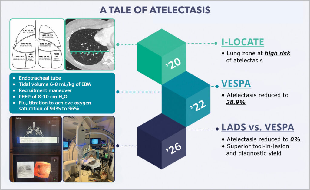
Published in 2020, the I-LOCATE (Incidence and Location of Atelectasis) trial was an observational prospective study in which 57 patients undergoing bronchoscopy under general anesthesia were subject to an atelectasis survey carried out with R-EBUS at the end of bronchoscopy. Eight bronchial segments in dependent areas (right bronchus [RB] 2, RB6, RB9, RB10, and left bronchus [LB] 2, LB6, LB9, LB10) were assessed for atelectasis. At a median time of 33 minutes from anesthesia induction to atelectasis survey, 89% (95% CI, 78%-96%) of participants had atelectasis in at least one bronchial segment. The highest incidences occurred in dependent lung segments, with rates of 70% to 79% in LB6, LB10, and RB10. Not surprisingly, risk factors for atelectasis include higher BMI and longer procedural time.

To characterize by chest CT scan the areas of the lungs with increased risk of atelectasis, in a post hoc analysis Khan and colleagues studied the CBCT images obtained during the VESPA (Ventilatory Strategy to Prevent Atelectasis) trial.4 To simplify their findings and provide our readers with a practical tip, if we draw a horizontal line at the anterior surface of the vertebral body in the CT slice corresponding to our target and our target is located posterior to this line, it is at high risk (greater than 50%) of being obscured by atelectasis. It is in these cases that a preventive strategy should be used, particularly in patients with high BMI and lower lobe lesions.
Effective strategies to mitigate atelectasis
Ventilatory and positional strategies have been recently described. The VESPA trial was designed to prevent atelectasis during nodal sampling with a convex-probe endobronchial ultrasound (C-EBUS). Seventy-six patients undergoing C-EBUS were randomized 1:1 to a control group with standard ventilation (laryngeal mask airway, 100% Fio2 and zero PEEP) or VESPA group (endotracheal intubation; Fio2 titration as low as feasible to maintain oxygen saturation between 94% and 96%; PEEP between 8 and 10 cm H2O; tidal volumes [VT] of 6-8 mL/kg ideal body weight [IBW]; and a recruitment maneuver of 10 breaths at a plateau pressure of 40 cm H2O with 20 cm H2O of PEEP). The proportion of patients with any atelectasis (unilateral or bilateral) on CBCT scan performed 40 to 45 minutes from induction of anesthesia was reduced from 84% (95% CI, 72.6%-95.8%) in the control group to 29% (95% CI, 15.4%-45.9%) in the VESPA group (P < .001).
There were no significant differences between the two groups regarding complication rates, which were all minor; 24% of the control group experienced hypotension requiring intermittent doses of vasopressors compared with 29% of the VESPA group.5 Retrospective reports of local experiences with other ventilatory strategies, with VT and PEEP twice as high as those in VESPA, and with prolonged breath-holds—likely used to compensate for the exaggerated target motion arising in the very high VT they selected—also reduced atelectasis rates to the 20% to 30% range but caused hemodynamic instability in up to 70% of the patients.6
With the aim of fully eradicating atelectasis, the same group of investigators that carried out I-LOCATE and VESPA trials developed a positional strategy for robotic bronchoscopy, the Lateral Decubitus Strategy (LADS).7 This approach involves placing patients in lateral decubitus position with the target lesion side up, along with standard ventilation, without the need for PEEP or recruiting maneuvers. Their initial report on this strategy was followed by a randomized controlled trial of LADS vs VESPA comparing these two approaches in patients undergoing robotic bronchoscopy for nodules measuring 3 cm or smaller located in dependent lung zones (at-risk zone as described previously).8 Twenty-nine patients were randomized to LADS and 33 to VESPA. The median BMI was 27.9 (IQR 23.8-32.7), the median target size was 1.6 cm (IQR 1.1-2.1), and 87.1% of the targets were in the lower lobes. The primary outcome was atelectasis obscuring target in CBCT, which was reduced from 27.3% (95% CI, 13.3%-45.5%) in the VESPA group to 0% in the LADS group (P = .002).
Additionally, the LADS group had higher tool-in-lesion confirmation (100% vs 72.7%, P = .002) and higher diagnostic yield (86.2% vs 57.6%, P = .013) compared with that of the VESPA group. Patients who crossed over from the VESPA arm to the LADS arm after developing atelectasis obscuring targets experienced complete resolution of atelectasis, with subsequent tool-in-lesion confirmation in all cases and diagnostic confirmation in 78% of crossover patients. Procedure times, radiation exposure, and complication rates (only minor) were comparable between groups. LADS was found to be a highly successful strategy to prevent and eradicate atelectasis, leading to better procedural outcomes.8
The use of prone positioning (PP) to prevent atelectasis has been retrospectively described in small case series.9 While there is little doubt that PP would be effective (interventional radiologists commonly use PP for CT-guided percutaneous approach), it is evidently more complicated than LADS. PP requires an experienced team (typically not available in the bronchoscopy suite) to proceed with extreme caution and coordination. If any complication should arise (pneumothorax, bleeding, extubation, intravenous line dislodgement), these would all be harder to manage. Unlike in LADS where the target lung is free of atelectasis (shift of atelectasis to the dependent lung), in PP, atelectasis is shifted ventrally within the target lung, which can still cause CT-to-body divergence, being detrimental to navigational bronchoscopy.
Based on the available literature, LADS, when feasible, is the procedure of choice for patients with high BMI and lesions located in dependent areas.
Incorporating these considerations effectively
With diagnostic accuracy, patient safety, and procedural efficiency in mind, we recommend a systematic approach to robotic bronchoscopy. A reasonable strategy that targets the vital components of the preparation for robotic bronchoscopy is the so-called A, B, C, D strategy.10 In a nutshell, this includes atelectasis prevention, artificial airway selection and airway clearance, bed adjustment and patient positioning on the bed, C-arm isocentering and collision check, and docking of the robot. Estimating the risk of atelectasis obscuring target (based on patient’s BMI and target location), communicating with the anesthesia team, and selecting the most suitable prevention strategy, when needed, are cornerstone aspects of this approach.10
This article was originally published in the Fall 2026 issue of CHEST Physician.
References
1. Zeng C, Lagier D, Lee JW, Vidal Melo MF. Perioperative pulmonary atelectasis: part I. Biology and mechanisms. Anesthesiology. 2022;136(1):181-205. doi:10.1097/ALN.0000000000003943
2. Sagar AS, Sabath BF, Eapen GA, et al. Incidence and location of atelectasis developed during bronchoscopy under general anesthesia: the I-LOCATE trial. Chest. 2020;158(6):2658-2666. doi:10.1016/j.chest.2020.05.565
3. Kammer MN, Heideman BE, Maldonado F. Should we start with navigation or endobronchial ultrasound bronchoscopy? Insights from Monte Carlo simulations. Chest. 2022;162(1):265-268. doi:10.1016/j.chest.2022.02.040
4. Khan A, Bashour S, Sabath B, et al. Severity of atelectasis during bronchoscopy: Descriptions of a new grading system (Atelectasis Severity Scoring System “ASSESS”) and at-risk-lung zones. Diagnostics (Basel). 2024;14(2). doi:10.3390/diagnostics14020197
5. Salahuddin M, Sarkiss M, Sagar AS, et al. Ventilatory strategy to prevent atelectasis during bronchoscopy under general anesthesia: A multicenter randomized controlled trial (Ventilatory Strategy to Prevent Atelectasis -VESPA- Trial). Chest. 2022;162(6):1393-1401. doi:10.1016/j.chest.2022.06.045
6. Bhadra K, Baleeiro C, Patel S, et al. High tidal volume, high positive end expiratory pressure and apneic breath hold strategies (lung navigation ventilation protocol) with cone beam computed tomography bronchoscopic biopsy of peripheral lung lesions: results in 100 patients. J Bronchology Interv Pulmonol. 2024;31(2):105-116. doi:10.1097/LBR.0000000000000938
7. Lin J, Sabath BF, Sarkiss M, Jimenez CA, Casal RF. Lateral decubitus positioning for mobile CT-guided robotic bronchoscopy: a novel technique to prevent atelectasis. J Bronchology Interv Pulmonol. 2022;29(3):220-223. doi:10.1097/LBR.0000000000000844
8. Boster JM, Goertzen M, Sarkiss M, et al. Superiority of lateral decubitus strategy in preventing atelectasis from obscuring targets during robotic bronchoscopy: lateral decubitus strategy vs ventilatory strategy to prevent atelectasis trial. Chest. 2026;169(4):1124-1134. doi:10.1016/j.chest.2025.11.044
9. Alraiyes AH, Johnson C, Madjer N, et al. Prone positioning in cone beam CT-guided robotic bronchoscopy case series: A strategy to minimize atelectasis and improve access to posteromedial lower lobe nodules. J Thorac Dis. 2025;17(11):9275-9286. doi:10.21037/jtd-2025-1413
10. Casal B, Casal RF. Best practices in shape-sensing robotic bronchoscopy with mobile cone beam computed tomography guidance: how I do it. J Thorac Dis. 2026;18(2):168. doi:10.21037/jtd-2025-1-2507
