
Imaging
-
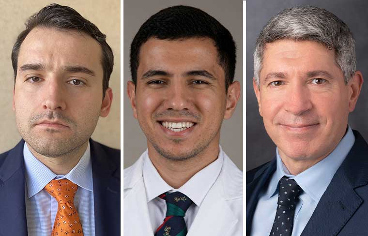
Atelectasis in peripheral bronchoscopy
Atelectasis remains one of the greatest challenges during robotic bronchoscopy, reducing diagnostic accuracy and obscuring peripheral lung lesions. Learn how evidence-based ventilatory and positioning strategies can minimize atelectasis, improve tool-in-lesion confirmation, and enhance procedural success.
-
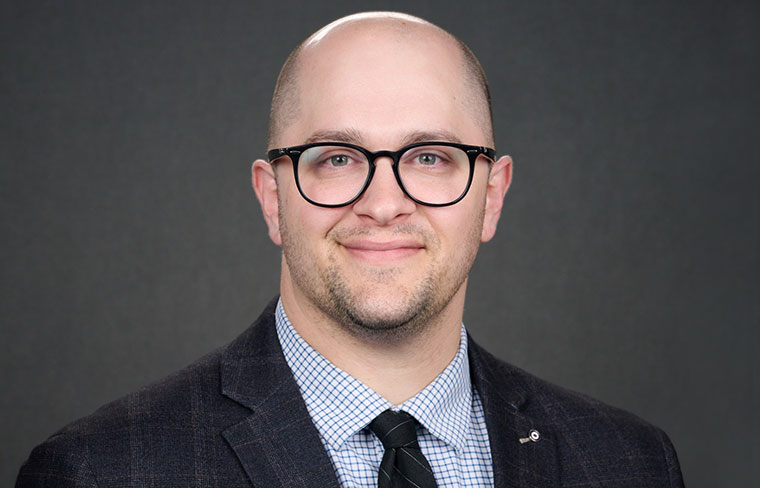
Advancing diaphragm dysfunction care
Diaphragm dysfunction is frequently underrecognized despite its significant impact on breathing and quality of life. Learn how an APP-led specialty clinic uses POCUS, standardized assessment, and physician collaboration to improve diagnosis, streamline care, and expand patient access.
-

The evolution of interventional pulmonology
Interventional pulmonology has rapidly evolved into a technology-driven specialty, with advances like robotic bronchoscopy and real-time imaging enabling safer, more precise access to peripheral lung lesions. These innovations allow clinicians to combine diagnostic and therapeutic procedures in a single setting.
-

AI-enabled approaches poised to transform diagnosis, management of lung diseases
Experts explore how AI-based tools can be used to bridge diagnostic challenges and address unmet needs in chronic respiratory disease care.
-

AI tools like Sybil poised to improve lung cancer screening, risk prediction
The Sybil model has shown promising results in predicting lung cancer risk in diverse populations from a single CT scan, which could address gaps and disparities in current screening guidelines.
-

AI applications in pulmonary medicine: From training clinicians to transforming diagnostics and treatment
AI applications in health care are multiplying, and AI-focused research is gaining traction across specialties, including pulmonary, critical care, and sleep medicine.
-

Imaging panel discusses pace, direction of practical AI applications
Amit Gupta, MD, MRMD, CIIP, spoke about the growing application of AI imagery in early-stage lung cancer detection.
-
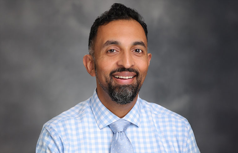
Radiologists, pulmonologists to discuss identification of interstitial lung abnormalities
Joe Mammarappallil, MD, PhD, will chair a Monday afternoon session on the recognition and risk stratification of ILAs, including how to differentiate imaging red herrings from findings that point toward progressive fibrosis.
-
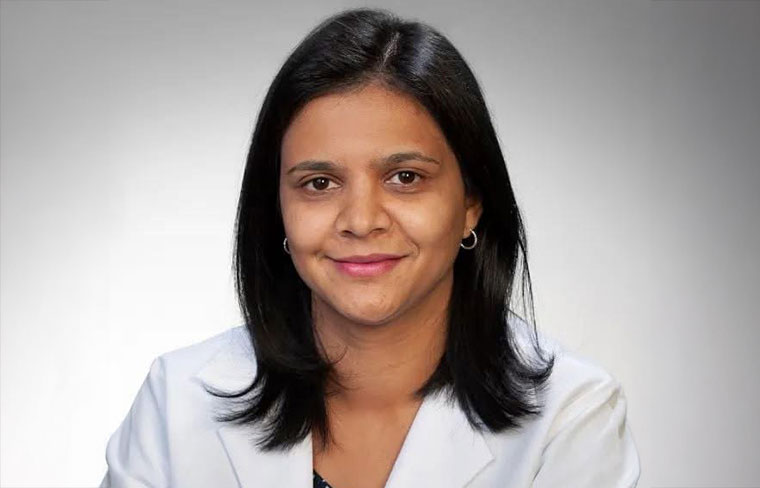
Experts to explore imaging modalities for the often-overlooked right heart
Navitha Ramesh, MD, MBBS, FCCP, will chair a session on available imaging techniques and considerations for evaluating the complex structure and function of the right ventricle.
-
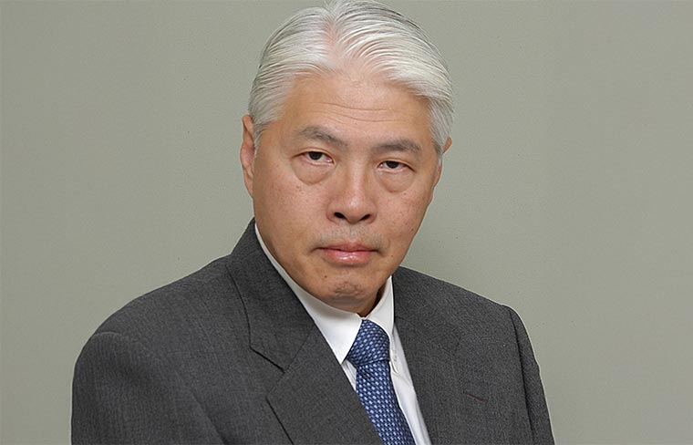
Updates in ninth edition of TNM classification criteria allow for more precise lung cancer staging
Hisao Asamura, MD, said the changes are critical to clinical decision-making in the evolving thoracic oncology treatment landscape.
